Medically reviewed by Dr Goutham Sivasuthan Specialist Surgeon, MBBS, MSurg, BSc, Co-Founder and CEO of Precursor. AHPRA MED0002000354. Last reviewed 8th September 2026.
Cost: $2000 - $3500
Medicare: Not covered
Time: About 1 hour
Radiation: None
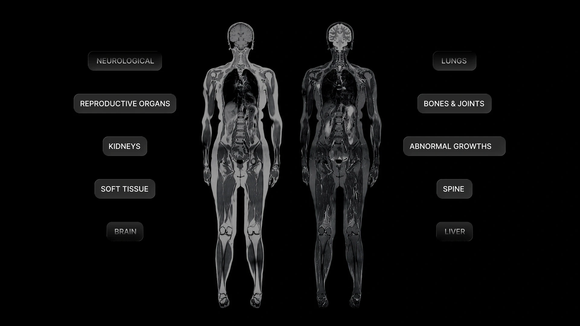
A full-body MRI creates detailed images across several areas without ionising radiation. Used thoughtfully, it can add a valuable structural view to a longer-term health plan.
Many people already track blood results, fitness or family history. MRI contributes something different: a broad set of images that a radiologist can review for structural changes. The result can help you and your clinician decide whether anything deserves a closer look.
Why full-body MRI appeals in proactive health
A full-body MRI brings several body regions into one imaging appointment. That breadth is useful for people who want to understand more about their health and prefer to make considered decisions before a problem becomes disruptive.
MRI is non-invasive and does not use ionising radiation. It is particularly good at showing many soft-tissue structures, including organs in the abdomen and pelvis. In some settings, MRI can show abnormalities that are less visible with other imaging methods. (1, 3)
The scan creates a structural overview rather than a score. A radiologist reads the images and prepares a report. That report becomes another layer of information for a clinician to place beside your health history, blood results and established screening needs.
What can a full-body MRI show?
MRI uses magnetic fields and radio waves to create detailed images inside the body. A full-body protocol collects images across several regions, while a targeted MRI concentrates on one organ or a specific clinical question. (1)
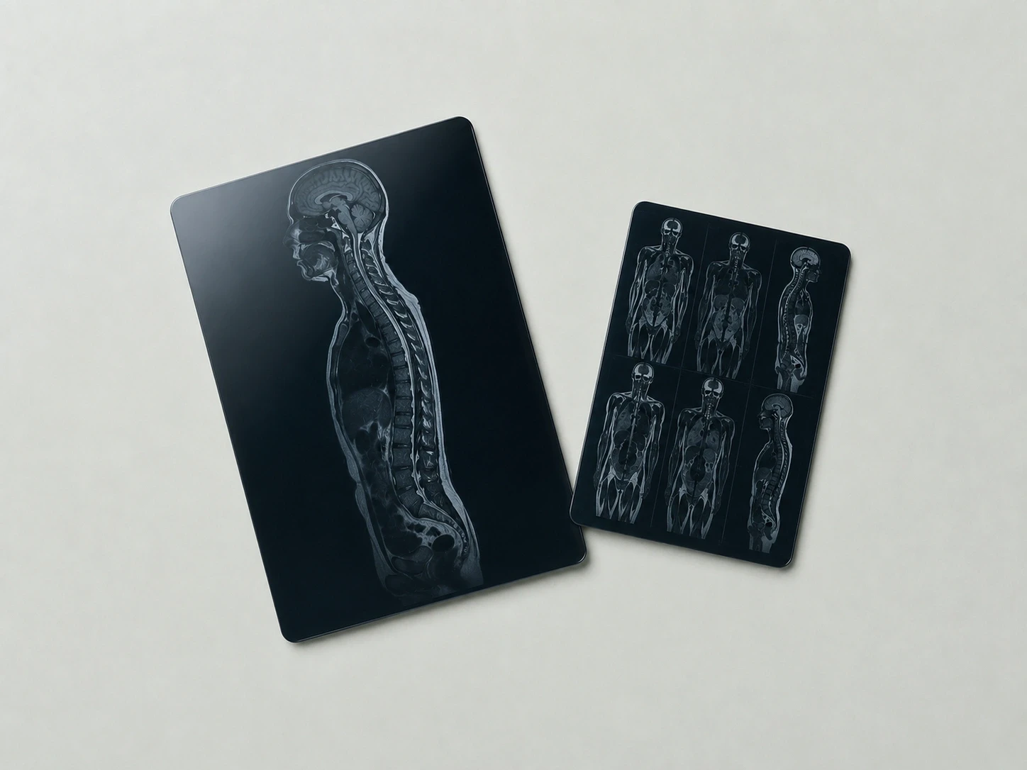
The images may identify a change that deserves further assessment. Published screening studies have reported findings involving organs, blood vessels, the spine and other tissues. The breadth of the scan is part of its appeal. (2, 7)
Protocols vary in coverage, imaging sequences and contrast use. This is why the result is best understood as a broad structural view rather than every possible dedicated examination combined into one appointment. A targeted scan may still be the better test for a particular symptom or organ. (2)
How MRI can fit a longevity plan
A longevity plan works best when information leads to a useful decision. Full-body MRI can contribute by documenting how structures look at one point in time and identifying findings that may warrant a focused review.
This structural information complements other parts of preventive care. Blood tests describe biochemical markers. Physical assessments show function and fitness. Established cancer-screening programs use tests designed for particular populations and conditions. MRI adds imaging to that wider picture. (8)
The value comes from clinical context. A finding may support follow-up, while a reassuring report can document what was visible within the scan’s scope. Either result is more useful when someone who understands your health history explains what it means and what comes next.
A calm and comfortable scan experience
During the examination, you lie on a padded table that moves into the MRI scanner. You remain still while the images are collected. The machine is noisy, so hearing protection is provided, and the imaging team can communicate with you throughout the scan. (1, 3)
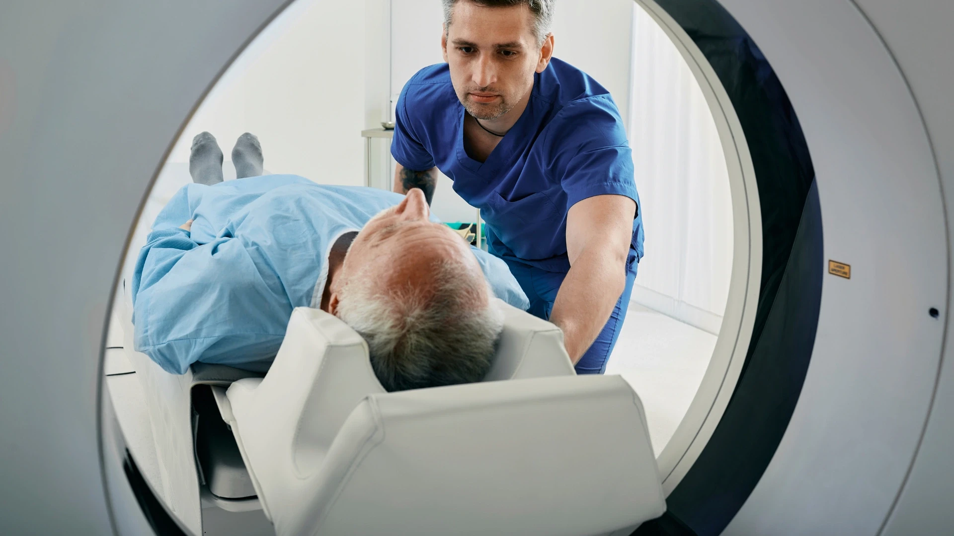
MRI itself is painless. Appointment length depends on the protocol and preparation. People who feel uncomfortable in enclosed spaces can discuss support before the day. If sedation or medicine for anxiety is used, the imaging team will provide transport and driving instructions. (3)
The referral process also adds a useful clinical checkpoint. In Australia, MRI is generally arranged through a doctor’s referral. This gives the examination a documented reason and connects the report to someone who can interpret it in context. (3)
Turning the report into a useful next step
An unexpected finding does not automatically mean something is wrong. Some changes are harmless, while others benefit from targeted imaging or a specialist opinion. A planned review helps separate findings that need attention from those that do not. (2, 5)
This is where a supported pathway matters. A radiology report contains technical descriptions. The clinician reviewing your results can translate those descriptions into a proportionate plan, taking account of symptoms, prior imaging and your broader risk profile.

False positives, false negatives and overdiagnosis are recognised features of screening. They do not remove the potential value of imaging. They explain why the report should be interpreted rather than treated as a pass-or-fail verdict. (5, 6, 7)
What a proactive scan can and cannot promise
A full-body MRI can provide detailed structural information. It cannot show every disease, measure blood chemistry or guarantee future health. A normal report therefore means that no concerning finding was reported within the examination’s scope.
For people without symptoms, a previous cancer diagnosis or a cancer-predisposition syndrome, the Royal Australian and New Zealand College of Radiologists does not recommend routine whole-body MRI screening.
People with inherited cancer risk or an established surveillance plan are in a different situation. Their imaging should continue under the guidance of the treating team. New or persistent symptoms also need their own clinical assessment, even after a recent screening scan.
Australia’s breast, bowel, cervical and lung cancer-screening programs remain important for people who meet their eligibility criteria. A full-body MRI can sit beside appropriate preventive care; it does not replace these programs. (8)
Safety and preparation
The imaging team screens every person for implanted devices, previous operations and possible metal fragments. Many implants can be scanned under defined conditions. Exact device details help the team plan safely. (1)
Pregnancy and possible pregnancy should be disclosed before imaging. A clinically needed MRI and an elective screening scan involve different decisions, and non-urgent imaging may be postponed. (1)
Some examinations use a gadolinium-based contrast injection. When contrast is part of the protocol, the imaging team considers its purpose alongside kidney health and any previous contrast reaction. The benefits and risks are discussed before it is given. (9)
Preparation varies. The instructions supplied for the appointment cover food, medicines, clothing and arrival time. If a CT or another X-ray-based examination is included separately, that component uses ionising radiation even though the MRI does not. (3, 10)
Full-body MRI with Precursor
Precursor places imaging within a broader, clinician-guided view of health. The aim is to help you understand what the scan adds, how it connects with your other results and which next step is worth taking.
Explore Precursor’s full-body MRI service to review the current pathway and see how the scan may fit your goals. You can proceed with a clearer sense of what it offers and how the result could contribute to longer-term health planning.
This information is general and does not replace advice from your treating doctor.
TAKE CONTROL OF YOUR HEALTH
Find the precursor, shape the outcome.
Sources
1.
2.
4.
5.
6.
7.
8.
9.
Frequently asked questions
What are the main benefits of a full-body MRI?
A full-body MRI produces detailed images across several body regions in one examination. It is non-invasive and does not use ionising radiation. The scan may identify structural changes that deserve closer assessment, giving you and your clinician another useful layer of information for longer-term health planning. (1, 2)
How can a full-body MRI support a longevity plan?
Full-body MRI adds a structural view to information from blood tests, health history and physical assessments. It may identify a finding that changes the next step or provide a documented baseline within the scan’s scope. MRI has not been shown to extend life in average-risk people, so clinical interpretation remains important. (2, 4, 5)
Can a full-body MRI detect cancer?
A full-body MRI may identify some cancers or findings that need targeted assessment. It cannot detect every cancer or provide a universal all-clear. Established cancer-screening programs and symptom-led investigations still have distinct roles. The report is most useful when a clinician interprets it alongside your risk profile. (2, 7, 8)
Does a full-body MRI use radiation?
No. MRI uses magnetic fields and radio waves rather than ionising radiation. This makes it well suited to detailed soft-tissue imaging without X-ray exposure. Safety screening is still needed for implants and metal. Any separate CT component does use ionising radiation. (1, 10)
Can I consider full-body MRI if I feel well?
Yes, you can discuss it as an elective, self-funded choice with a clinician. The expected value depends on your health history, risk and what the result would add. Australian and US radiology colleges do not recommend routine screening for average-risk people because a survival benefit has not been established. (4, 5)
Do I need a referral for a full-body MRI in Australia?
MRI in Australia is generally arranged through a doctor’s referral. The referral connects the scan to a clinical reason and gives the radiologist relevant context. It also creates a clear pathway for discussing the report and deciding whether any follow-up is useful. (3)
What is the full-body MRI experience like?
You lie still on a table while the scanner collects images. The examination is painless, although the machine is noisy and the enclosed space can feel unfamiliar. Hearing protection and communication with the imaging team are provided. Support can be planned in advance if you feel claustrophobic. (1, 3)
What happens after a full-body MRI?
A radiologist reviews the images and prepares a report for the referring clinician. The clinician explains any findings in the context of your health and decides whether monitoring, targeted imaging or another review would add value. Many incidental findings do not require treatment. (2, 3)